Mitosis And Meiosis
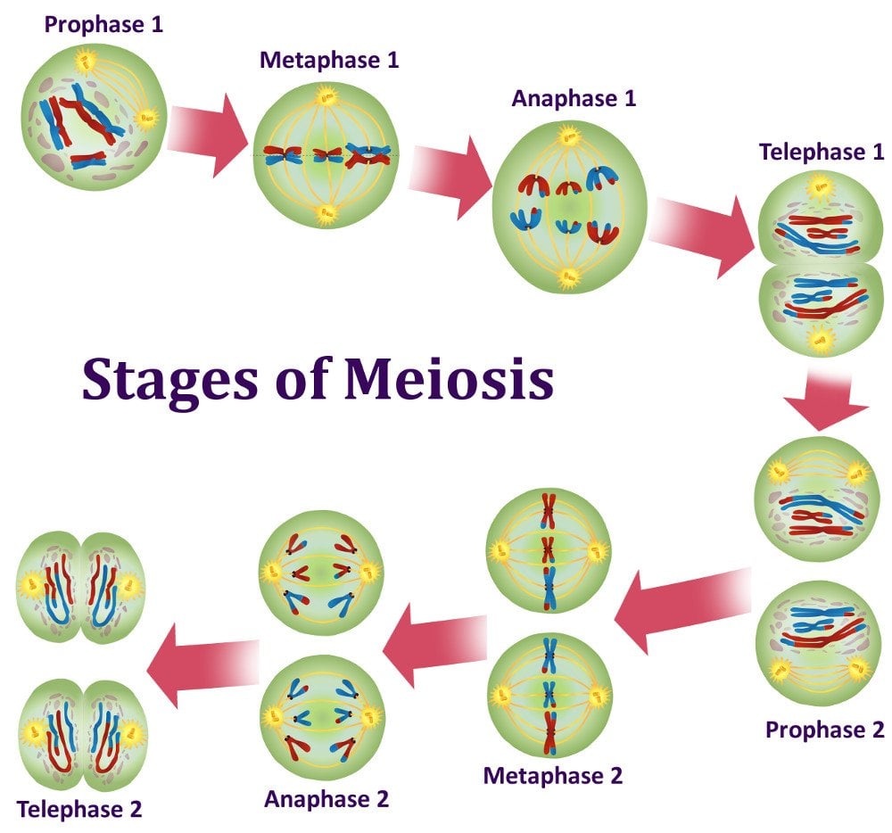
Need help with your assignment?FIND A WRITER OR TUTOR TO GRADE YOUR ESSAYMeiosis is a method where a solitary cell splits twice to generate four cellular material containing 1 / 2 the original quantity of genetic information. These kinds of cells will be our love-making cells – sperm in males, ova in females. During the process of meiosis a single cell splits two times to form four little girl cells.These several daughter cellular material only have fifty percent the number of chromosomes of the mother or father cell that happen to be called haploids. Meiosis makes our sex cells or perhaps gametes which can be (eggs in females and sperm in males). Meiosis can be split up into nine stages. These are divided between the first time the cell divides (meiosis I) plus the second period it divides (meiosis II): Meiosis I. Interphase: First, the GENETICS in the cellular is replicated resulting in two identical total sets of chromosomes.
In that case. Outside of the nucleus happen to be two centrosomes with each containing a couple of centrioles, these kinds of structures happen to be critical for the process of cell department. During the process of interphase, microtubules extend via these centrosomes. Prophase I: The copied chromosomes shortened into X-shaped set ups that can be conveniently seen within microscope. Then simply every other chromosome is composed of two sister chromatids having the same genetic details. The chromosomes pair up so that both copies of chromosome one are jointly, both clones of chromosome two are also together.
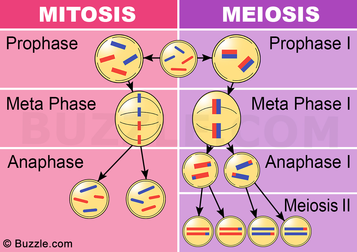
Mitosis and Meiosis. Is the process of nuclear cell division. It is the process of chromosome segregation and nuclear division that follows replication of the genetic material in eukaryotic cells. The main difference between mitosis and meiosis is that mitosis is the type of cell division which takes place in somatic cells for growth or for asexual reproduction in some organisms while meiosis is the type of reproduction which takes place in sex cells for the intention of sexual reproduction.
The pairs of chromosomes will then interchange chunks of GENETICS in a process called recombination or traversing over. At the end of Prophase one, the membrane about the nucleus in the cell dissolves away, liberating the chromosomes. The meiotic spindle, comprising microtubules and also other proteins, extends across the cellular between the centrioles.
Metaphase I: The chromosome pairs line up next to each other over the center which can be the (equator) of the cell. The centrioles are now at opposites poles of the cell with the meiotic spindles elevating from them. Then a meiotic spindle fibers attach to one chromosome of each pair. Anaphase I: Then this pair of chromosomes are pulled apart by the meiotic spindle, that pulls one chromosome to 1 pole of the cell as well as the other chromosome to the opposite pole.
In meiosis 1, the sister chromatids stay together which is different from what happens in mitosis and meiosis II. Telophase I actually and cytokinesis: The chromosomes finished their particular move to the contrary poles from the cell. At each pole in the cell, an entire set of chromosomes get together.
A membrane varieties around in every set of chromosomes to create two new nuclei. Then solitary cell pinches in the middle to form two individual daughter cellular material in which every single contains an entire set of chromosomes within a center.
This process may be cytokinesis. Meiosis 2. Prophase II: At this point, there are two child cells with 23 chromosomes each (23 pairs of chromatids).
In every single two child cells, the chromosomes condense again in to visible X-shaped structures that could be easily viewed under a microscopic lense. The membrane around the nucleus in every single daughter cellular dissolves away releasing the chromosomes. Then this centrioles replicate and the meiotic spindle forms again. Riders of icarus gameplay. Metaphase II: In each one of the two daughter cells the chromosomes (a pair of sister chromatids) get in line end-to-end along the equator of the cell. Right now the centrioles are at opposites poles in each of the daughter cells. Meiotic spindle fibers at each rod of the cellular attach to each one of the sister chromatids. Anaphase II: The sister chromatids are after that pulled to opposite poles due to the actions of the meiotic spindle.
The separated chromatids are in that case individual chromosomes. Telophase II and cytokinesis: Finally the chromosomes complete their particular move to the opposite poles with the cell. At each pole of the cell, a full set of chromosomes get together. A membrane varieties around in every set of chromosomes to make two new cell nuclei. This can be the last phase of meiosis, however, cell division remains not finish without an additional round of cytokinesis.
When cytokinesis can be complete there are four granddaughter cells, every single with half a set of chromosomes (haploid): in males, these types of four skin cells are all semen cells in females, among the cells is an egg cellular while the different three will be polar physiques which are (small cells which experts claim not develop into eggs).Mitosis: Mitosis is the process where a eukaryotic cell nucleus separates in two, and then division of the parent cell into two daughter skin cells. The word “mitosis” means “threads, ” and it identifies the thread-like appearance of chromosomes since the cell prepares to separate. These tubules, collectively referred to as spindle, increase from buildings called centrosomes, with 1 centrosome located at each with the opposite ends, or poles, of a cellular.
As mitosis starts advancing, the microtubules join towards the chromosomes, that have already replicate their DNA and have get together across the middle of the cell. The spindle tubules then simply contract and move toward the poles of the cell. As they maneuver, they move the one backup of each chromosome with these to opposite poles of the cell. This process makes sure that each child cell is going to contain one particular exact replicate of the parent cell GENETICS.What Are the Phases of Mitosis?Mitosis is composed of five morphologically distinct levels: prophase, prometaphase, metaphase, anaphase, and telophase. Each one of these stages involves attribute steps in the process of chromosome alignment and separating.
Once mitosis is full, the entire cellular divides in two by the process named cytokinesisProphase which is the initial stage in mitosis, happening after the summary of the G2 portion of interphase. During prophase, the father or mother cell chromosomes, which were replicated during T phase — condense and turn into thousands of moments more compact than they were during interphase. Due to the fact that each copied chromosome consists of two the same sister chromatids joined for a point known as the centromere, these buildings now seem as X-shaped bodies once viewed under a microscope. Several DNA joining proteins catalyze the condensation process, including cohesin and condensin. Cohesin forms rings that hold together the sis chromatids, whereas condensin varieties ring that coil the chromosomes in to highly compact forms.
Likewise, the mitotic spindle begins to develop during prophase. As the cell’s two centrosomes move against opposite poles, microtubules gradually assemble between them, forming the network which will later pull the duplicated chromosomes separate.What Happens during Prometaphase?After prophase is total, the cell enters prometaphase which is the other stage of mitosis. During prometaphase, phosphorylation of nuclear lamins simply by M-CDK triggers the nuclear membrane to break down into quite a few small vesicles.
Therefore, the spindle microtubules now have direct access to the innate material from the cell. Every single microtubule is highly dynamic, growing outward from the centrosome and falling straight down backward as it tries to choose a chromosome.
Eventually, the microtubules find their targets and hook up to each chromosome at its kinetochore, a complex of proteins located at the centromere. The actual number of microtubules that link to a kinetochore varies between species, but at least a single microtubule from each post link to the kinetochore of each and every chromosome. A tug-of-war after that ensues since the chromosomes move back and forth between the two poles.What goes on during Metaphase and Anaphase?Since prometaphase ends and metaphase begins, the chromosomes change along the cell equator.
Just about every chromosome has at least two microtubules extending from the kinetochore and with for least one particular microtubule attached to each post. At this point, the strain within the cell becomes well-balanced, and the chromosomes will no longer push back and forth. Additionally, the spindle is now total, and three groups of spindle microtubules happen to be apparent.
Kinetochore microtubules hook up the chromosomes to the spindle pole, intercalar microtubules develop from the spindle pole over the equator, nearly to the opposing spindle rod, and astral microtubules develop from the spindle pole to the cell membrane. Metaphase then leads to anaphase, during which every single chromosome sibling chromatids individual and go on to opposite poles of the cellular. Enzymatic breakdown of cohesin — which will linked the sister chromatids together during prophase — causes this kind of separation to occur. Upon separation, every chromatid becomes an independent chromosome. Changes in microtubule span provide the system for chromosome movement. Specifically, in the 1st part of anaphase — occasionally called anaphase A — the kinetochore microtubules cut short and attract the chromosomes against the spindle poles. After that, in the second part of anaphase — at times called anaphase B — the spirit microtubules that are anchored to the cell membrane pull the poles additional apart plus the interpolar microtubules slide past each other, exerting an additional take on the chromosomes.

During telophase process, the chromosomes arrive at the cell poles, the mitotic spindle disassembles, plus the vesicles that include fragments in the original nuclear membrane assemble around the two sets of chromosomes. Phosphatases then dephosphorylate the lamins at every end of the cellular. This dephosphorylation results in the introduction of a new elemental membrane around each band of chromosomes.Remedy Cells Actually Divide?Cytokinesis is a physical method that finally splits the parent cell into two identical child cells. During this process, the cell membrane layer pinches in at the cellular equator, building a cleft called the cleavage furrow. The position from the furrow will depend on the position with the astral and interpolar microtubules during anaphase.
The boobs furrow in that case forms due to action of your contractile ring of overlapping actin and myosin filaments. As the actin and myosin filaments move transferring each other, the contractile ring becomes smaller, akin to drawing a drawstring at the top of a handbag. When the engagement ring reaches their smallest level, the boobs furrow entirely bisects the cell in its center, leading to two separate daughter cells of the same size.
Mitosis is a process of asexual reproduction in which the cell divides in two producing a replica, with an equal number of chromosomes in each resulting diploid cell.
Meiosis is a type of cellular reproduction in which the number of chromosomes are reduced by half through the separation of homologous chromosomes, producing two haploid cells.
Following are the differences between Mitosis and Meiosis:
S.N. | Differences | Mitosis | Meiosis |
| 1 | Type of Reproduction | Asexual | Sexual |
| 2 | Genetically | Similar | Different |
| 3 | Crossing Over | No, crossing over cannot occur. | Yes, mixing of chromosomes can occur. |
| 4 | Number of Divisions | One | Two |
| 5 | Pairing of Homologs | No | Yes |
| 6 | Mother Cells | Can be either haploid or diploid | Always diploid |
| 7 | Number of Daughter Cells produced | 2 diploid cells | 4 haploid cells |
| 8 | Chromosome Number | Remains the same. | Reduced by half. |
| 9 | Chromosomes Pairing | Does Not Occur | Takes place during zygotene of prophase I and continue upto metaphase I. |
| 10 | Creates | Makes everything other than sex cells. | Sex cells only: female egg cells or male sperm cells. |
| 11 | Takes Place in | Somatic Cells | Germ Cells |
| 12 | Chiasmata | Absent | Observed during prophase I and metaphase I. |
| 13 | Spindle Fibres | Disappear completely in telophase. | Do not disappear completely in telophase I. |
| 14 | Nucleoli | Reappear at telophase | Do not reappear at telophase I. |
| 15 | Steps | Prophase, Metaphase, Anaphase, Telophase. | (Meiosis 1) Prophase I, Metaphase I, Anaphase I, Telophase I; (Meiosis 2) Prophase II, Metaphase II, Anaphase II and Telophase II. |
| 16 | Karyokinesis | Occurs in Interphase. | Occurs in Interphase I. |
| 17 | Cytokinesis | Occurs in Telophase. | Occurs in Telophase I and in Telophase II. |
| 18 | Centromeres Split | The centromeres split during anaphase. | The centromeres do not separate during anaphase I, but during anaphase II. |
| 19 | Prophase | Simple | Complicated |
| 20 | Prophase | Duration of prophase is short, usually of few hours. | Prophase is comparatively longer and may take days. |
| 21 | Synapsis | No Synapsis | Synapsis of Homologous chromosomes takes place during prophase. |
| 22 | Exchange of Segments | Two chromatids of a chromosome do not exchange segments during prophase. | Chromatids of two homologous chromosome exchange segments during crossing over. |
| 23 | Discovered by | Walther Flemming | Oscar Hertwig |
| 24 | Function | Cellular reproduction and general growth and repair of the body. | Genetic diversity through sexual reproduction. |
| 25 | Function | Takes part in healing and repair. | Takes part in the formation of gametes and maintenance of chromosome number. |